SHERLOC (Scanning Habitable Environments with Raman and Luminescence for Organics and Chemicals)
mission specific
Instrument Overview
SHERLOC will assess the habitability potential of an ancient site including understanding its aqueous history. Identification of clay minerals, carbonates, sulfates, and halides in sedimentary units has shown that Mars hosted nearly a dozen different types of aqueous (alkaline, acidic, and saline lakes, hydrothermal deposits etc.) and other potentially habitable environments during its first billion years. Some minerals are particularly indicative of environmental conditions such as serpentine (alkaline, reducing), sulfate (acidic, oxidizing), and carbonates (alkaline). Other phases are useful as environmental indicators though only when they are found in mineral assemblages where multiple phases collectively allow derivation of fluid geochemistry. By collecting point mineralogical data over an extended spatial scale in conjunction with texture, it is possible to discern whether aqueous minerals formed in sediments, hydrothermal precipitates, volcanic materials, or resulted from secondary overprinting by weathering or diagenesis. Measurements on these scales, achievable by SHERLOC can identify many important features including evidence of aqueous activity, through analysis of clasts, alteration amygdules, sedimentary laminae, melt pockets, or individual grains in conglomerates or breccias. SHERLOC measurements on the micron to multi-millimeter scale are an important size-step in the continuum of contextual investigations that make up the 2020 mission.
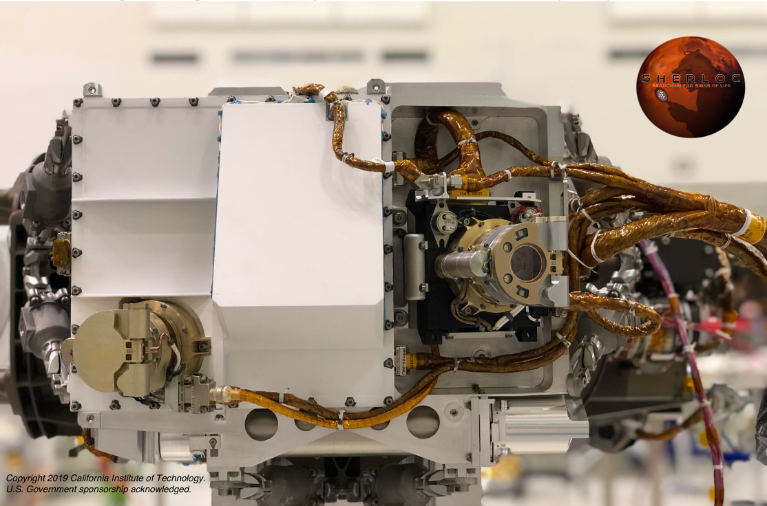
The SHERLOC Turret Assembly on the Perseverance robotic arm during the ATLO test with both spectroscopic ACI boresight (left) and WATSON boresight (right) visible.
SHERLOC would assess the availability of key elements and energy source for life. Life on Earth is driven by oxidation-reduction reactions that form and transform organic matter and mineral substrates containing key elements such as C, H, N, O, P and S. The abundance and diversity of these substrates in a sedimentary environment are key measures of habitability. Analysis of these substrates in a given environment can identify sedimentary rocks that reflect the detrital component, a precipitated residue of the dissolved component, and post-depositional (diagenetic) alteration processes. Transformation of minerals by biology or diagenesis has distinctive signatures that can be observed in assemblages of astrobiologically important minerals (e.g., carbonates, nitrates, phosphates, sulfates, etc.). The presence of such minerals, especially in association with organics, or when they otherwise imply some degree of chemical or morphologic disequilibrium, is an important component in the evaluation of potential biogenicity.
SHERLOC will ascertain if there are potential biosignatures preserved in Martian rocks and outcrops. Potential biosignatures that can be identified and spatially resolved are key organics such as hopanes, steranes, organic macromolecules, etc. This especially strong interrogative power will help identify samples that will facilitate making the case for returning them to Earth. SHERLOC's macro/micro-mapping modes can determine the organic and hydrated minerals present over scales that match the mineralogy and morphology of most biosignatures resulting from microorganisms. It is capable of correlating detected classes of organics with morphology (widths and shapes) to determine whether morphological candidates for microfossils, filaments, or stromatolitic layering are potentially biogenic. SHERLOC is a robotic arm-mounted, Deep UV (DUV) native fluorescence and resonance Raman spectrometer. It utilizes a DUV laser to generate characteristic Raman and fluorescence photons from a targeted spot. The DUV laser is co-boresighted to a context imager and integrated into an autofocusing/scanning optical system that allows us to correlate spectral signatures to surface textures, morphology and visible features. The context imager has a spatial resolution of 30 µm and currently is designed to operate in the 400-500 nm wavelength range.
Through the use of an internal scanning mirror, autofocusing lens, and a depth of focus of ±250 µm, the 50 µm laser spot can be systematically scanned over a 7x7 mm area with a fine-scale spatial resolution on natural or abraded surfaces and boreholes to a depth of ~25 mm, without further arm movement. Through the use of the context imager, SHERLOC's data products can be combined with observations made by other instruments on the Mars 2020 payload [3]. By bringing to bear multiple scientific instruments on a single sample, our ability to assess the habitability of ancient environments and search for potential biosignatures preserved within the geologic record will be greatly enhanced, making possible the selection of high-priority samples for caching. The SHERLOC investigation combines two spectral phenomena, fluorescence and pre-resonance/resonance DUV Raman scattering. These spectral features are resolvable when a high-radiance, narrow line-width, laser source illuminates a sample. In fluorescence, the incident photons are absorbed and re-emitted at a longer wavelength. The difference between the excitation and emission wavelength is the difference between the excitation frequency and the lowest electronic state frequency that increases with increasing aromatic structure (i.e., number of aromatic rings). Typical fluorescence cross-sections are 107x greater than traditional Raman, enabling the detection of sub-picograms levels of aromatic organic compounds.
Fluorescence emission of organics extends from ~270 nm into visible wavelengths. On the other hand, mineral fluorescence emission stemming from crystalline defects and impurities is weak in the deep UV, and typically begins longward of 360 nm continuing through the visible and into the NIR. Mineral fluorescence is very unlikely to be seen in samples found on Mars as the only reported fluorescence of naturally occurring inorganics in the region 250–360 nm is in non-relevant astrophysical conditions. In over 30 years of experiments in our laboratories, the only observed fluorescence at shorter wavelengths (<360 nm) have always been directly attributable to organics trapped inside the mineral matrix of a field sample, not the mineral itself. Thus, the DUV fluorescence technique employed by SHERLOC is well-suited to the detection of organics on mineral surfaces.
SHERLOC's narrow-linewidth (<3 GHz) DUV laser (248.6 nm) also enables fluorescence-free resonance/pre-resonance Raman scattering for additional classification of aromatics and aliphatic organics, and minerals. Raman scattering is inelastic scattering involving loss of photon energy corresponding to vibrational energy. Raman shifts (cm-1) (i.e., peak position as photon energy loss from the excitation energy) are invariant to excitation wavelength thus peak positions can be compared with values in in Raman databases for all excitation wavelengths (including 229, 248, 488, 532 and 785 nm).
As stated, traditional Raman scattering cross section are roughly 107 times smaller than the corresponding fluorescence cross-sections. The strength of the induced Raman signal is dependent on incident laser power, laser wavelength and resonance effects. Rayleigh Law states that the intensity of the Raman scattered light has an intensity µ l-4, resulting in 20x greater scattering efficiency at 248.6 nm, than at 532 nm and 100x greater than at 785 nm. Adding to the increases in signal strength, many organics have resonance and pre-resonance signal enhancements of 100 to 10,000 times when excited by 248.6 nm. Finally, many astrobiologically relevant minerals have the same pre-resonance effects at these excitation wavelengths, increasing the Raman signal without high power lasers (see figures below).
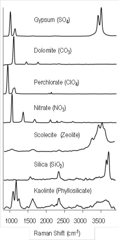
Example Raman spectra of astrobiologically relevant minerals.
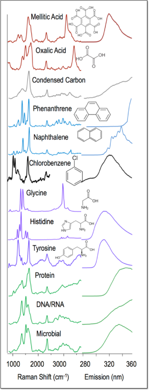
Example Raman and Fluorescence spectra of some important organic molecules.
In order to identify Raman spectral features, it is necessary to block the Rayleigh scattered incident laser light. The laser is injected into the optical path by a Semrock edge-filter that performs within specifications over the range of Martian temperatures. This filter rejects wavelengths <253 nm including the high intensity Rayleigh scattered from the excitation source. This corresponds to a frequency interval between the laser and lowest Raman frequency of 0 to 810 cm-1 (248.6 and 253.7 nm). SHERLOC targets organics and mineral Raman spectral features that exist above of 810 cm-1 and includes organic functional groups features corresponding to C-H, CN, C=O, and C=C bonds, and carbonates, perchlorates, sulfates, and phyllosilicates. These functional groups and minerals represent the highest priority astrobiological target species.
Excitation at DUV wavelengths (<250 nm) enables resonance and pre-resonance signal enhancements of between 100 and 10,000 times of organic/mineral vibrational bands. As such, SHERLOC combines the detection of both the Raman and fluorescence photons on the same CCD for simultaneous detection. The spectrometer CCD and related electronics have the necessary detector dynamic range to enable detection of both spectral features simultaneously. In addition, the lack of a background fluorescence in DUV Raman spectral region, greatly simplifies the resulting analysis. The figure below shows an example observation of a stromatolite. The chert signature observed is highly attenuated by the edge filter but is observable in this sample. Colors blend in the map and organics are green and yellow as they are mixed with the chert signature (sample courtesy of A. Allwood).
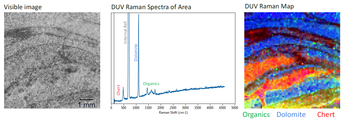
Example of SHERLOC science on the Stelley Pool Stromatolite using the lab breadboard MOBIUS instrument [2][5].
SHERLOC Spectroscopic Investigation Matrix
The table below lists Mars 2020 mission goals and derived Mission Science Objectives, mapped into SHERLOC contributions and Data Products addressed by the SHERLOC investigation.
| Derived Mission Science Objectives |
SHERLOC Analysis |
SHERLOC Data Product |
Notes |
|---|---|---|---|
| Mars 2020 Objective A: Decipher geological processes and assess past habitability | |||
|
Assess how long aqueous conditions existed and identify organic mineral assemblages that constrain chemical and redox environment) |
Classify mineral assemblages associated with aqueous activity. |
DUV Raman spectra of carbonates, sulfates, phyllosilicates, zeolites, etc. with grain sizes >50 μm. |
DUV Raman signatures of astrobiologically important organic and mineralogical species describe energy sources, ancient environmental conditions and water availability at a time when the surface of Mars was warmer and wetter. |
|
Assess availability and distribution of electron donors, key elements and energy sources |
Detect and classify CHNOPS elements in minerals and organics. Identify alterations in mineral content due to biologic processes. |
DUV Raman spectra of CHNOPS minerals in organics/minerals over an area at 50 μm resolution. |
|
| Mars 2020 Objective B: Assess the biosignature potential preservation and search for potential biosignatures | |||
|
Characterize physiochemical processes and conditions in the paleoenvironment, and identify mechanisms for formation and preservation of biosignatures |
Classify organics within mineralogical context and assess the probability of origin by meteoritic infall, native, and/or biologically derived processes. |
Textural, fluorescence maps of organics & DUV Raman maps of organics and minerals. Includes grain size analysis (>50 μm) and distribution. |
Combining fluorescence and DUV Raman signatures for bulk and fine scale organic and mineral maps of surface/nearsurface material describes the potential for organic preservation and potential biosignatures detection. Combining mineral and organic maps enables in situ assessment of formation and alteration processes for a given target to assess whether materials are meteoritic, native and/or past life and chemical precursors. Also constrains the potential for biosignature preservation. |
|
Characterize degree, type, and timing of diagenetic processes that could have degraded biosignatures. |
Assess thermal, oxidation, hydroxylation, etc., via organics and mineralogy analysis. |
DUV Raman Maps of organics and minerals. |
|
|
Map distributions of potential biosignatures (PBS) relative to geologic features, including characteristics of potentially biogenic structures |
Assess organic classes (aromaticity, functional groups, PBS, etc.) and correlating aqueous-related mineralogy and textures on surfaces and boreholes. |
DUV Raman and fluorescence maps of organics and minerals co-located with textures and morphology of the target surface over a 5 x 5 mm area with spatial resolution of 100 μm. Borehole mapping of mineral and organic species to 25 mm depth. |
|
| Mars 2020 Objective C: Future return of scientifically selected, well documented samples to Earth | |||
|
Assess evidence for past life, or its chemical precursors, and place constraints on the past habitability and the potential for preservation of the signs of life |
Assessment of mineral/ textural characteristics consistent with PBS, energy sources, or preservation. Identification of preserved evidence suggesting biotic or prebiotic signatures. |
DUV Raman and fluorescence maps of organics and minerals co-located with textures and morphology of the target surface over a 5 x 5 mm area with spatial resolution of 100 μm. Borehole mapping of mineral and organic species to 25 mm depth. |
Facilitates merger of organic/mineral data with other payload instruments to improve analytical capability of the rover instrument suite. Identification of samples with organic composition is vital to selection of samples for return. The direct analysis of boreholes results in a proxy for core analysis. SHERLOC's mapping mode inside the borehole can identify organic gradients that indicate degradation of organic material. |
|
Assess the history and significance of surface modification processes including but not limited to: impact, photochemical, volcanic, and aeolian. Constrain the magnitude, nature, timing and origin of past planet wide climate change |
Assess morphologic and mineralogical characteristics of Mars' paleoenvironment with co-located organic molecule mapping, mineral phase mapping, and contextual images. Identify salt, aqueous products, and evaporite concentrations. |
Deep UV Raman and fluorescence maps of organic molecules co-located with perchlorates or other oxidizers and textures, (grain size, shape, distribution) co-located with surface features. |
|
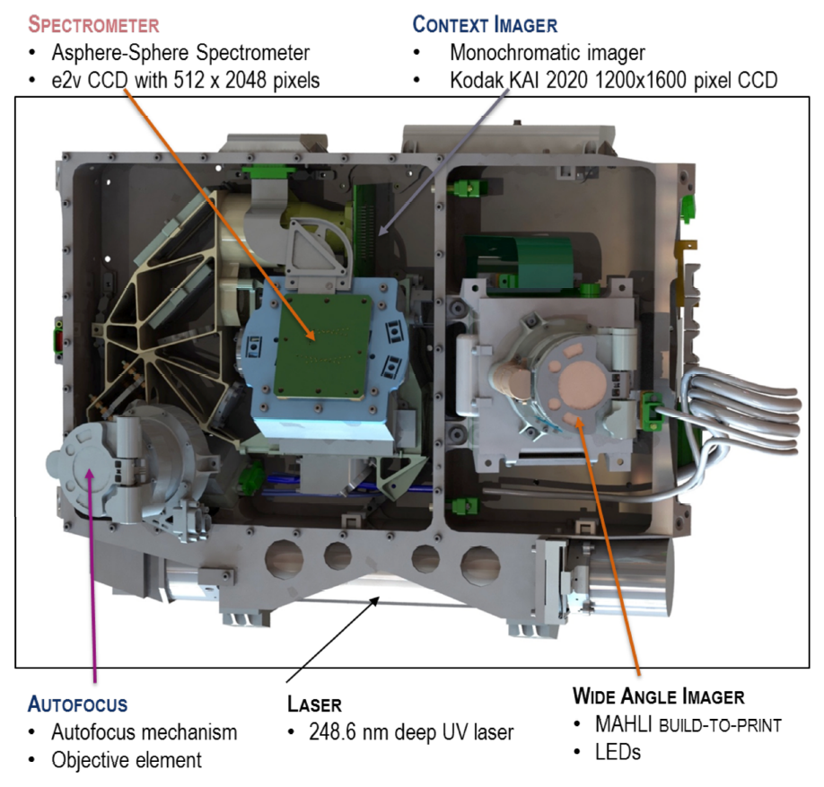
Instrument Functional Description
The SHERLOC instrument is an arm-mounted, deep ultraviolet (DUV) native fluorescence and resonance Raman spectrometer and imager. SHERLOC uses a DUV laser to illuminate the target and generate the characteristic Raman and fluorescence spectral response. SHERLOC contains two imaging subsystems: Autofocus and Context Imager (ACI) and Wide Angle Topographic Sensor for Operations and eNgineering (WATSON). The DUV laser is co-boresighted with the ACI and an integrated scanning optical system that allows correlation of spectral signatures to surface textures, morphology and visible features. Through the use of an internal scanning mirror, autofocusing lens, and a depth of focus, the SHERLOC laser spot can be systematically scanned over the spectroscopy map area with a fine-scale spatial sampling on natural or abraded surfaces and borehole interior walls, without further arm movement. Through the use of the context imager, SHERLOC's data products will be combined with the other instruments on the Mars 2020 payload. This allows greatly increased scientific analysis by bearing down the entire scientific measurements on a single sample to assess the habitability of ancient environments and search for potential biosignatures preserved within the geologic record to select high-priority samples for caching.
General Science
The SHERLOC investigation will enable spatially resolved, high-sensitivity detection and characterization of organics and minerals in the Martian surface and near subsurface. SHERLOC's goals are to assess past aqueous history, detect the presence and preservation of potential biosignatures, and to support selection of return samples. To do this SHERLOC will detect CHNOPS-containing minerals, detect the distribution and type of organics preserved at the surface, and correlate them to textural features.
Measurement Descriptions
SHERLOC's investigation combines two spectral phenomena, native fluorescence and pre-resonance/resonance DUV Raman scattering. These events are resolvable when a high-radiance, narrow line-width, laser source illuminates a sample. In fluorescence the incident photon is absorbed and re-emitted at a higher wavelength. The difference between the excitation wavelength and the emission wavelength is correlated with the number of electronic transitions between excitation and emission which increases with increasing aromatic structure (i.e., number of aromatic rings). Typical cross-sections are 105x greater than Raman, enabling the detection of sub-picograms of carbon. Native fluorescence emission of organics extends from ~270 nm into the visible. Conversely mineral fluorescence emission stemming from crystalline defects and impurities does not have strong absorption features in the deep UV resulting in mineral fluorescence that typically begins longward of 360 nm and continues through the visible into the NIR. The only reported fluorescence of naturally occurring inorganics in the region 250–360 nm is in non-relevant astrophysical conditions. In over 30 years of experiments in our laboratories, the only observed fluorescence at shorter wavelengths (<360 nm) have always been directly attributable to organics trapped inside the mineral matrix of a field sample, not the mineral matrix.
SHERLOC's narrow-linewidth (3 GHz) DUV laser (248.6 nm) also enables fluorescence-free Raman scattering for additional classification of aromatics and aliphatic organics and minerals. Raman scattering is inelastic scattering where the loss of energy from the excitation energy (40225 cm-1) and defines the vibrational energy of a bond with which it interacted. This enables classification of bonds such as C-H, CN, C=O, C=C, NHx, NOx, SOx, POx, ClOx, and OH.
Calibration Target
In addition to the two imaging subsystems and a DUV laser spectrometer, the Mars 2020 rover carries an external SHERLOC Calibration Target (SCT). The SCT is affixed to the front of the rover and includes ten calibration targets to enable instrument calibration on the Mars Surface.
The figure below captures the ten calibration targets and their location within the calibration target palette.
The calibration target material is being identified as:
| Target | Material |
|---|---|
| 1 | AlGaN on Sapphire Raman Region (~262 nm peak) |
| 2 | Diffusil Diffuser (Bubbled Silica) |
| 3 | Mars Meteorite SaU 008 |
| 4 | Intensity Target (Chromium on Silica) |
| 5 | AlGaN on Sapphire Fluorescence Region (~335 nm peak) |
| 6 | Polycarbonate Over Opal Glass Geocache |
| 7 | Vectran Fabric |
| 8 | Orthofabric |
| 9 | Teflon Fabric |
| 10 | nGimat Coated Teflon Fabric |
SHERLOC camera suite
The SHERLOC instrument has two cameras, WATSON and ACI. WATSON is used for general observations while ACI captures details of the spectrometer observation. . The figure below shows the locations of both SHERLOC cameras
WATSON (Wide Angle Imager) and ACI (Context Imager) Cameras of SHERLOC instrument suite.
SHERLOC-WATSON
The SHERLOC-WATSON camera is a focusable color camera located on the turret at the end of the M2020 robotic arm and is a build-to-print copy of the MSL MAHLI instrument. The instrument acquires images of up to 1648 by 1200 pixels (generally only 1600 x 1200 is used) with a color quality equivalent to that of consumer digital cameras using a Bayer pattern. It is also capable of video. SHERLOC-WATSON optics characteristics useful in the analysis of EDR and RDR products are described in the table below. Note that the spatial resolution in measures the working distance. Working distance is measured from the front lens element (front surface of sapphire window) to the target (assumes a planar target parallel to the plane defined by the sapphire window). Working distance can be determined from focus motor count per the formulae (for dust cover close and dust cover open cases) in the Edgett et al. (2019) WATSON Cal Rept.
| SHERLOC-WATSON Operational Characteristics | |
|---|---|
| Characteristic | Value |
| Resolution (S x L) | 1648 x 1200 |
| Bit Depth | 11 |
| Field of View (FOV) | close range focus = 41° infinity focus = 39° |
| Spatial Resolution |
~105 μm/pixel at 27 cm distance 15.9 μm/pixel at 25 mm distance |
| Angular Resolution | 0.3 - 0.34 mrad/pixel |
| Spectral Wavelength ± Bandwidth (λeff ± HWHM) | 590 ± 88 nm (Broadband) |
| 640 ± 44 nm (Bayer filter Red) | |
| 554 ± 38 nm (Bayer filter Green) | |
| 495 ± 37 nm (Bayer filter Blue) | |
| Focal Length | 18.3 - 21.3 mm |
| f/number | 9.8 - 8.5 |
| Depth of Field | 1.6 mm - >4800 mm |
| Focus Range | 17.8 mm - infinity |
| Number of Spectral Filters | 1 plus Bayer pattern on CCD |
Spatial resolution may be calculated by:
PS = 6.7593 + [3.658 × WD]
where PS is the Pixel Scale in μm/pixel and WD is the working distance is in cm. Note that the constants above are actuals for the unit onboard the M2020 Spacecraft, and these differ from testbed-WATSON, testbed-MAHLI, and MSL-flight-MAHLI).
Autofocus and Context Imager (ACI)
The SHERLOC-ACI is used to capture detailed images of the area observed by the SHERLOC spectrometer. It is co-boresighted with the laser illumination and spectral collection path. Light from the target (indirect sunlight or LED illumination) is collected by the autofocus objective and directed to the context imager by the foreoptics. The table below provides the camera characteristics.
| SHERLOC-ACI Operational Characteristics | |
|---|---|
| Characteristic | Value |
| Resolution (S x L) | 1648 x 1200 |
| Bit Depth | 11 |
| Field of View (FOV) | 7.9 x 10.6 degrees 16.16 x 12.12 mm with 11.7 mm ⌀ unvignetted |
| Spatial Resolution | 10.1 μm/pixel at 48 mm distance |
| Angular Resolution | 115 μrad/pixel |
| Spectral Wavelength | 550 ± 50 nm (grayscale) |
| Depth of Field | ± 250 μm |
| Number of Spectral Filters | 0 (monochrome detector) |
| Effective focal length | 87.5 mm |
| Working distance | 48 mm |